

In a significant advancement for cancer treatment, researchers have developed a groundbreaking artificial intelligence (AI) tool capable of detecting lung tumors with exceptional accuracy—tracking their movement with every breath a patient takes.
The new system, called iSeg, is the first 3D deep learning segmentation tool designed to map tumors as they shift during respiration, offering oncologists a powerful new asset in their mission to deliver precise and effective radiotherapy.
Tackling a Critical Challenge in Radiotherapy
Radiotherapy demands precision. Before targeting tumors with high-dose radiation, oncologists must define the exact size and location of cancerous growths. This process, known as tumor segmentation, is traditionally done manually—an approach that is both time-consuming and prone to variation between physicians. As a result, areas containing cancer may sometimes go undetected, potentially compromising treatment outcomes.

3d rendered medical illustration of male anatomy – lung cancer. plain black background. anterior view.
The iSeg tool, developed by a team at Northwestern Medicine in collaboration with the Feinberg School of Medicine at Northwestern University, addresses this challenge head-on. Unlike previous AI systems that rely on static images, iSeg operates in real time, accounting for the movement of tumors with each breath—a critical factor in lung cancer treatment.
Outperforming Manual Detection
According to research published in Precision Oncology, iSeg was trained using CT scans and manually segmented tumor outlines from hundreds of lung cancer patients treated at Northwestern Medicine facilities and the Cleveland Clinic. When tested on new patient scans, iSeg matched doctors in its ability to outline tumors—but went further by identifying additional cancerous areas that had been missed by human experts.
These hidden regions are often linked to poorer outcomes if left untreated. By catching them, iSeg has the potential to significantly enhance the effectiveness of cancer care. As lead researcher Dr. Mohamed Abazeed, professor of Radiation Oncology at Feinberg, explained that we are one step closer to cancer treatments than we could have imagined just a decade ago.

Expanding to Other Cancers and Imaging Methods
While initially developed for lung cancer, the research team is now testing iSeg in real-time clinical environments and exploring its application to other types of tumors, including those of the liver, brain, and prostate. The tool is also being adapted to interpret results from other imaging technologies such as MRIs and PET scans, broadening its potential impact.
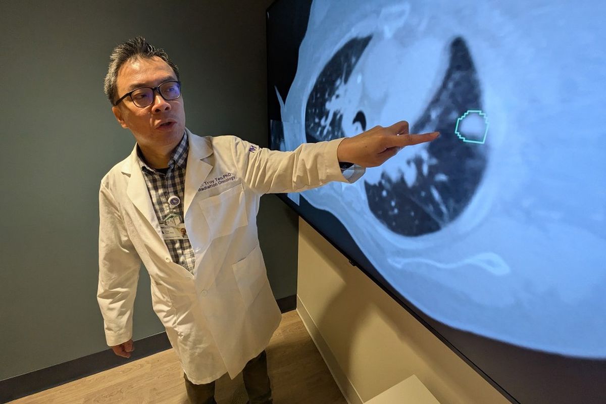
Dr. Troy Tao, another principal author of the study and professor of Radiation Oncology at Feinberg, emphasized the importance of this innovation, particularly in settings with limited access to specialized medical professionals. He noted that this tool, they hope, will improve targeting precision in radiation oncology, adding that widespread clinical implementation could be possible within the next one to two years.
The Future of AI in Oncology
The development of iSeg marks a critical step forward in the integration of artificial intelligence into routine cancer care. By enhancing accuracy in tumor detection and supporting more personalized treatment plans, it represents a leap toward a future where technology and medicine work hand in hand to improve survival rates and patient outcomes.